Category Archives: The SCIENCE

Spinal anatomy, biomechanics of setting & paleo-anthropology

Disc Nutrition & Spinal Movement.

Spinal movement & change of position are important for a number of reasons including IV disc nutrition.

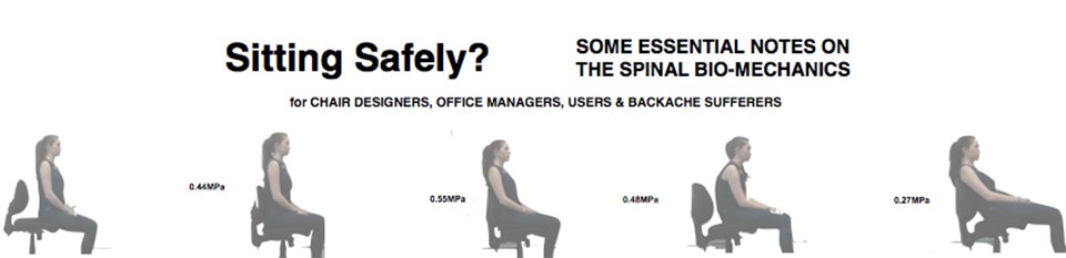
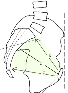
Intervertebral Disc Nutrition starvation plays a part in nuclear degeneration.  Nutrients are only barely supplied to the central part of the Intervertebral disc by diffusion from surrounding blood vessels in the circumference and the endplates .  Fluid flow also plays a part, especially for the transport of the larger molecular metabolites, and is dependent on the pumping action of pressure changes of the compression/decompression that occur with movement, such as changing from a supine to upright posture (Wilke H-J, 1999.).    Some disc narrowing occurs rapidly but is then followed further, as water is forced out over a longer timescale, and is near maximal in about 6 hours.  This is evident in diurnal variation when the torso is 15-25 mm longer (and stiffer) in the morning when intra-discal fluid pressure is at its highest. Fluid is expelled during the day, while standing, when the compressive force exceeds the osmotic pressure force (Adams MA, Hutton WC.).     During recumbency the disc regains 71% of it’s height in the initial 3.75 hrs but with 3 hrs of upright posture 80% of height loss has occurred.  This accounts for some of the early morning stiffness experienced by those with minor degenerative disc changes and also for the change in height of astronauts on their return from a weightless environment.

A prolonged static position is uncomfortable.  This should be distinguished from supported static ‘comfort’ which is much sought after by chair manufacturers but may not necessarily be  biomechanically sound.   On the other hand, what is biomechanically sound is always comfortable.

Spinal movement has a ‘comforting’ effect so that movement is enjoyed .  This is due to physiological effects and is slightly different from the rhythmic rocking chair movement which was recommended to President Kennedy for his back pain which has a slightly different method of action depending on pain relief by afferent sensory bombardment of the central nervous system.

Although difficult to quantify in terms of sitting and the design of chairs, it is important to arrange for changes in posture, from reclining to upright, to occur frequently during the day to allow changes in axial loading to benefit disc nutrition.   It also allows the user the comfort of movement and helps to avoid the discomfort of point pressure in badly designed systems.   Respiration is also said to be helped by avoiding a prolonged cramped posture.  General wellbeing, for example, in aiding blood flow and venous return, with other health risks avoided (Aaras, 200045).  Rani Lueder, a respected American ergonomist and researcher, reviews the subject of spinal movement and comes down firmly in support of movement based chairs(200245}.  Gorman (201051), on the other hand, remarked “However the history of chairs with lots of movement is that people don’t like them and they die out pretty quickly…… Also if, as I suggest, pumping and movement is not the central point of low back pain then lots of the justification for such a chair is gone.  I have always argued that it is the flexion of the lowest two discs beyond the natural limit that is the central problem. (48 & see his URL → )

Sensors integrated into the surface of the seat can alert users who have not changed their position in a long time (.Dr. Thomas Hermann with the Ambient Intelligence research group).

For an account of remediation see ☛ EXERCISE & movement

In the context of the 2Tilt concept  movement is at the users command by entering  the stable end-range modes.  See  ☛The unstable TRANSITIONAL MODE. It’s importance.


How did it all begin?  A good start is with the pelvis of  A afarensis, an individual known as ‘Lucy’.  

Photo on 06-12-2013 at 15.17

Australopethecine bipedalism has resulted in considerable morphological changes to the pelvis when compared to that of other apes.   An australopithecine pelvis. Similar to that of a human.Screen Shot 2013-09-26 at 22.08.53

  1.  Ilium
  2.  SI Joint
  3.  Sacrum 
  4.  hip joint
  5.  pubic symphysis
  6. Ischial tuberosity

The pelvis of A. afarensis required rotation of the sacrum to increase the Lumbo-sacral wedge angle.   Lucy’s ilium (Togue, Lovejoy 198664) is similar to that of humans,  being short and having widely flared iliac blades.

Screen Shot 2013-09-14 at 22.11.29This morphological change is due to the shortening of the pelvic ilium.  The iliac blades are rounder and the outer surface face postero-laterally to give an attachment for the origins of the gluteal muscles.   These therefore have changed their function from hip extensors to become abductors and lateral rotators of the hip.  and are important for stabilising the pelvis on the hip and act to support the weight of the torso at the phase of walking when the contralateral, other leg is off the ground. A failure of this mechanism results in the pelvis tilting downwards away from the affected side and is the basis of the clinical Trendelenberg test. Non-hominid primates, with glutei only acting as hip extensors, therefore have an ambling gait or what doctors would describe as a Trendelenberg gait in a human.


The early hominids, such as Homo erectus, had a brain of 900 cc. and its primitive variant, of 1.8 MYA, found at  the Dmanisi (Georgia) site was only 650-780cc. These were probably the earliest hominids outside of Africa.    H. sapiens, with a volume of about 1300 cc appeared about 130,000 years ago according to the previous ‘out of Africa’ theory’.

The obstetric pelvis

 Lovejoy (Tague, Lovejoy 1986, 2005) pointed out, that the enlarging human brain (cerebralisation) in the foetal head required morphological changes of the pelvis to allow an adequate birth passage and changes of the lower half of the pelvis, composed of the ischium and pubis.
Pelvis of A afarensis The shortening of the ilium, an effect of bipedalism, results in the consequent unwanted effect of reducing the antero-posterior (AP) diameter of the pelvis and the size of the birth canal.  Although this was no great problem for early hominins, such as ‘Lucy’, as they had ape sized heads, it presents a problem in parturition for the larger brained hominids.      With the shortening of the ilium,  the distance between the hip joint and the Screen Shot 2013-09-14 at 23.32.19sacro-iliac (SI) joints is reduced and the sacrum lies at a lower position in relation to the pubic symphysis.  The human cranium is x4 larger than Australopithecine ape, and needs a larger pelvic AP diameter and an increase in width (coronal dimension) and lordotic curvature of the sacrum.    In the diagram :- X. True’ pelvic inlet of H.sap .     Y. Outlet     Z. The mid-plane conjugate diamete, the limiting dimension for childbirth was between “… the pubic symphysis and the centre of the sacrum”

In traversing the pelvic birth canal the human cranium has to engage at the inlet in the transverse plane.   In the mid-plane it rotates and change direction during it passage which is allowed by the sacrum becoming curved, with the radius of its arc centred at the pubis.  The head finally emerges in an AP plane at the outlet.

The pelvis of A. afarensis requires rotation of the sacrum which increases the wedge angle of each lower lumbar IV discs.

An obstetric dilemma, due to the increasing foetal encaphalisation, was increased by the changes to the pelvis and the lumbo-sacral junction due to the development of bipedalism.   Even in the best arranged modern facilities disproportion can occur between the maternal pelvis and the foetal head.  Jay Stock, at Cambridge (2011), has pointed out that a variety of other variable factors can also be involved, both for the size of the maternal pelvis and the foetal head resulting in disproportion, including ecological stresses such as the thermal environment and nutrition.   Maternal stature, obesity and increasing neonatal growth can be related to dietary shifts such as occurred with the development of agriculture.

next see ☛BIOMECHANICS

Screen Shot 2013-10-13 at 14.47.34


WHY? Mandal’s Homo sedens.

Screen Shot 2013-10-13 at 17.16.44AC Mandal was a leading Danish surgeon who wrote the    ‘The Seated Man’119.  Becoming disillusioned in the conventional wisdom of the principles of correct chair design, he transferred his scientific and critical training to that of the biomechanics of sitting.     ☞ Mandal

He observed that young children naturally sit with the chair tilted forwards onto the front legs.    They then support their elbows on the table, which for a small child is the right height, and the protective lumbar lordosis is effortlessly and correctly maintained.   As the child grows the height of the table becomes  relatively lowered and the child then sits in the usual semi slumped position. This results in short episodes of backache, which are ignored, and possibly to permanent stretching of the posterior spinal elements which can predispose to instability and a lifetime of adult  spinal pathology.
Screen Shot 2015-11-27 at 13.36.05

Drawing on the work of Keegan120 in the USA, Schorbath121 in Germany and Akerblom122 in Sweden he concluded that children are instinctively right to modify their seats to tilt forward with a slope of 10 to 20°. This allows the hip angle to open from 90° to 120° and  the pelvis to rotate forwards, on it’s fulcrum at the ischial tuberosities, so that the lowest two lumbar joints are in the extended (safe, lordotic) position, ensuring the correct lordotic posture.  No back rest was necessary and the torso was free to move.   His proposed solution was a combination of height adjustable desking and forward tilted seat similar to the equestrian seat.

The recommended ‘correct’ position was fundamentally wrong.    

Mandal
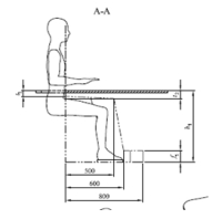
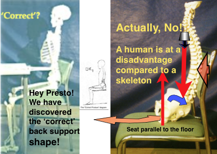
He suggests that this posture was developed by taking a standing skeleton and sitting it on a chair.

  • A skeleton has advantages over a live human.
  • It can sit motionless all day.
  • It has no constraining muscles ligaments or IV Discs.
  • It is steaded with an iron bar down the vertebral canal.


(The 4 (+2) main adverse effects apply :-

  • There is a x2.5 increase in spinal loading over full reclination on the discs and 40% over that of standing upright (Nachemson, Sato but not Wilke).
  • The pelvis tends to tilt backwards reducing the important and protective wedge (lordotic) angle at the lower 2 lumbar joints.
  • The position may be prolonged in an office or work environment.
  • Lumbar (not pelvic)  support accentuates this effect.
  • And the hip flexion also tilts the pelvis backwards.
  • the vulnerable lower lumbar joints are forced into a flexed position so that the protective high wedge angle is reduced to a point that the disc contents are liable to move backwards and protrude.
  •  See  BIOMECHANICS that determine safe sitting

Screen Shot 2018-12-20 at 14.46.36He wondered how such a poor position could have been accepted as correct.

  • A chapter in his book is titled ‘Functionalism’s instruments of torture’ and he dates the decline to the 1930 Exhibition in Stockholm which had the slogan “Beauty in everyday furniture”.   Later he met the authority who created this concept who admitted that the image of the back of a standing person was simply cut and pasted to a drawing of a sitting person (personal communication).  It has no scientific basis.  This basic false premise is still regarded as ‘correct’, regardless of more recent scientific knowledge of spinal pathology and bio-mechanics and is still universally advised with an air of authority.   Mandal claimed that 90% of lower back pain was related to loss of this lumbar lordosis.
  • With the increasing liability to backache, an ergonomically ‘correct’ chair was sought.
  • Wrongly!   ☛The upright seated posture.→ Screen Shot 2015-11-28 at 15.44.41

Screen Shot 2016-01-14 at 18.19.39       Google→ shows an amazing collection of diagrams and pictures of ‘correct’ seating with a few that are actually correct.   For a similar account see bodyzone  shops→

This mistaken view of correct seating is still accepted and widely promulgated by authorities who advise on such matters.  Some detailed examples  :-

Screen Shot 2017-02-11 at 17.18.18

Correct?

  1. Another diagram for correct seating actually spells out the arguably adverse ergonomic recommendations!  (outlined in red)

badsit5The lady is sitting  bolt upright, to attention, like a guardsman.  The body position shown is cramped and movement, which is necessary for comfort and disc nutrition, is hardly possible.

There is no armrest.  Some authorities maintain that this allows greater movement of the upper torso.  However support under the elbows prevents dropping of the shoulder girdle which can be tiring and uncomfortable.  Support at the wrists helps to prevent RSI.

The diagram shows the neck in the correct neutral position.  Unsupported cervical flexion results in much greater axial compression.  (This is equivalent to looking downwards when standing,  see Hansraj KK. 2014)

A greater viewing distance allows the intrinsic eye muscles to relax.   The rule should be to be as far from the screen as vision allows comfortably.  If necessary get glasses, tinted against glare, adjusted to 36 inches.

Screen Shot 2018-06-11 at 21.04.14
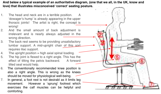
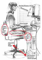
This diagram might be acceptable, a semi-partial remediation, if the point was made that it incorporated the alternative ‘Pelvic (iliac) support’ instead of ‘lumbar support’ as is shown. This alternative solution of applying the support directly to the pelvic (iliac) crest, ensures that it was unable to rotate backwards. This support should be a particular shape and size to spread the pressure loading.  (See later under ☛ Pelvic & Lumbar support→).

There are now many well engineered and comfortable office chairs in the market.      Screen Shot 2016-02-14 at 18.18.00Most have a fundamental bio-mechanical fault.  They rely on the mid- upright mode of sitting for prolonged work.  As has been shown this combines the two most adverse effects although the backward pelvic tilt can be modified by a FTS or lumbar support. My own opinion on a number of top range chairs, as shown, is of admiration of the design and engineering  mixed with awareness that the most essential bio-mechanical points have yet to be fully appreciated.   The following are better than most as efforts have been made to incorporate pelvic support (with reservations).Chairs2014-04-03 at 14.10.35


Ergonomics-standard-diagramThe latest version of BS EN 527-1: 2011,(the European standard specified dimensions for office desks and tables.)  state that fixed height desks should be 740mm ± 20 mm and desks should adjust between 650mm and 850mm for sitting modes and between 650mm and 1250mm for sit/stand desks.

The suggestion of a FTS would have been helpful.

Next, see ☛ Various chairs. How do they measure up?

Screen Shot 2013-10-13 at 14.47.34