The development of upright positional MRI (pMRI) scanners, allows imaging of the spine in the load-bearing postures which are relevant to sitting.
A pMRI study 2006 The first pMRI study of backward migration of the Nucleus Pulposus (NP) in functional positions was done at the Positional MRI Centre, Woodend Hospital, Aberdeen, U.K by Professor Francis Smith in 2006. For this study 11 symptom-free volunteers were recruited, aged between 18 and 60 years.
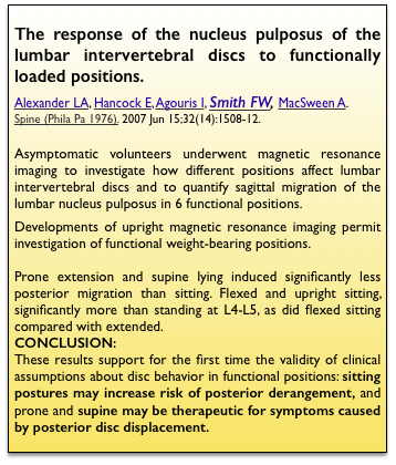
Results The authors concluded “These results support for the first time the validity of clinical assumptions about disc behavior in functional positions: sitting postures may increase risk of posterior derangement, and prone and supine may be therapeutic for symptoms caused by posterior disc displacement.” These results confirmed the views of JH Cyriax which he had published as far back as 19452 and subsequently by others3, ,4,5,6. It also validates the 2Tilt concept as the most effective, possibly the only, way to ensure ‘Safe Sitting’.
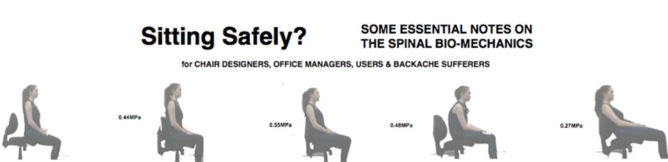
 The relevant weight bearing positions included sitting upright unsupported, with lumbar support (shown here) and slumped (also shown here). A standing position, which includes lumbar support is also shown here. Intradiscal pressure in this position was originally measured by Nachemson (1964) showed that intradiscal pressure to be 500-800 N for a 75 Kg man. See .
 Upright sitting, slumped.The usual position and was represented as 30% by students at Cambridge in a quick survey. Intradiscal pressure /load measured by Wilke (2001) was 0.48MP and by Sato 1127 kPa , 800N. This was less than sitting upright and can be explained by the pressure relieving effect of the abdominal cavity.
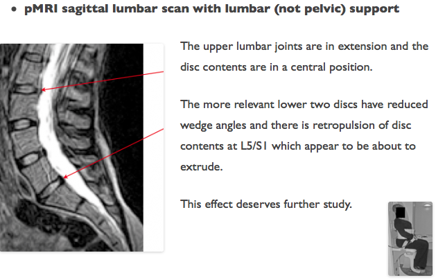
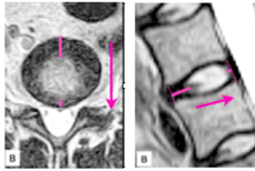
Upright sitting with lumbar (not iliac) support. As can be seen, the support is applied above the Iliac crest and can be expected to have an opposite effect at the lower 2 joints (Gorman). This is shown to be so.
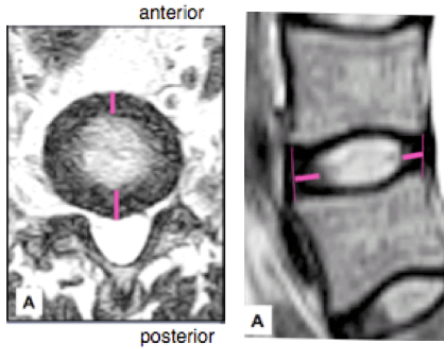
In the flexed sitting view (left), It can be seen that there is an adverse posterior position of the Nucleus Pulposus (NP) of the vulnerable L4/5 & L5/S1 discs. When compared to non-loaded, uncompressed disc scans (right), the NP has migrated, or in clinical terms there is retropulsion, to the position that is potentially dangerous.
 This is precisely what Cyriax had postulated in 1945.
Unexpectedly confirming the view postulated by JD Gorman the scan shows that support at the upper lumbar joints has a reverse, adverse, effect at the important lower joints. Lumbar v. pelvic support→ See Gorman’s view→
For a review from Working Ergonomics→
The pMRI evidence. Conclusion.
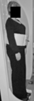
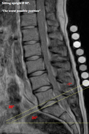
This investigation using Whole-body Positional MRI (pMRI), by FW. Smith, Bashir W (2007) found that the upright position, at 90°, caused disc contents to move the most, while the relaxed position (135°/45° reclined) caused disc contents to move the least. This confirms that the upright position is the worst for the back, while the relaxed position is the best. From which the following pictures are derived (arrows, etc, are added). This confirms the bio-mechanical evidence.
 pMRI scan in reclined, relaxed, sitting mode shows the NP in a safe mid-position. The hip angle is at 135°. the NP is in the safe mid-position. This is practical and preferable and is advocated for the 2Tilt principle in the reclined mode.
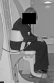
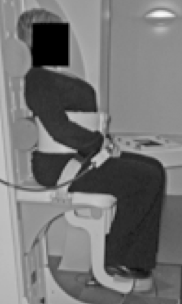
 pMRI scan in an upright sitting mode shows the NP has translated posteriorly which can culminate in protrusion. Hips are at an angle of 90° with the seat-pan horizontal.
This is visual confirmation of the bio-mechanical evidence.