Category Archives: The SCIENCE

Spinal anatomy, biomechanics of setting & paleo-anthropology

EXERCISE & movement

Exercise, spinal movement & change of position are important for a number of reasons including IV disc nutrition, for general health and for the avoidance of LBP.    

Effect of movement on the IV disc

An account of this can be seen at ☛ Disc Nutrition & Spinal Movement.→ .   It has been shown that disc nutrition depends on the pumping action of pressure changes due to changes of position and is probably important in avoiding later degenerative changes.  In the context of chair design, movement is comforting and avoids the adverse effects of a prolonged constrained static upright mode on general health (See (Lueder R 2002).

Effect of axial loading  on sitting

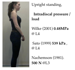
Originally Nachemson and others showed that the standing position had a lower intradiscal pressure than upright sitting.  This suggested that incorporating sitting and standing, with a sit/stand desk, could be beneficial if designed into an office environment.   This concept  has slowed following later work (Wilke 1999) showing less difference in sitting and standing intra-discal pressures than was first thought and the general recent ergonomic improvement of office chairs.    Most people prefer sitting, because it is energy efficient with less action by the Erector Spinae & leg muscles than when standing.

The Sit Stand concept

A comeback is occurring as the  importance of keeping staff exercised is recognised and  there are situations where the concept is applicable.   The Sit Stand concept is bio-mechanic superior to the prolonged use of most existing upright chairs and the negative consequences of constrained sitting which has been described by a number of authorities.   On the other hand, although it side-steps the issue of poor office seating it does not address the ergonomic requirements for prolonged work.


Screen Shot 2018-06-16 at 13.25.30

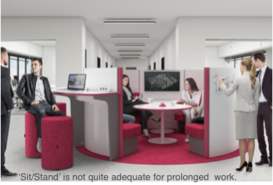
A ‘stand’ mode can  be incorporated into the 2T concept with a 3M ‘desk-less’ workstation.  This extra mode results in the  4M workstation, the first change since the inception of the concept in 1998 (and a ‘full’ solution).

Wobbly chairsScreen Shot 2018-09-22 at 14.05.14
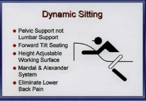
There is a recognition that prolonged constrained static postures are uncomfortable and deleterious for both spinal and general health.   Recently there has been interest in continuous small amplitude movement for upright chairs, the chair re-aligning with the users centre of gravity,  and termed  ‘Dynamic Seating’.     Exercise is required to maintain this position This provides proprioceptive feedback and frequent small amplitude pressure changes which may be comforting for short periods and helps multifidus muscle action. Rani  Lueder  gives a review account (Lueder R 2002) and the referenced evidence→ is considerable.

Screen Shot 2016-03-14 at 13.35.24

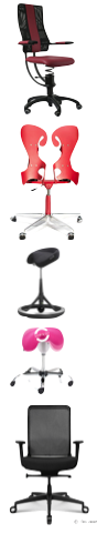
A few chairs are considered in this respect → various chairs. How do they measure up? .

Other ‘movement’  systems

Screen Shot 2015-11-21 at 17.15.57
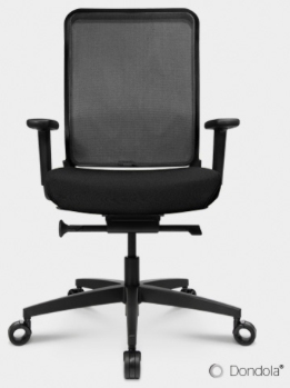
The Dondola hinge system allows a controlled all-round instability and a trial when incorporated in a Wagner chair showed reduction of morbidity when compared to the chair without the system.  However, there have been versions of floating, movable seats over the years with springs, rubber doughnuts, wobbly balls etc.  Not hugely commercially successful, they lacked stability in sitting and so were tiring and uncomfortable particularly for the legs which had to be used to maintain the stability that was not provided by the seat. This may be the same with the Dondola free float but the website is uninformative on the mechanism.   They say   “Nowadays we spend up to 14 hours in a seated position. 50% of all Germans suffer at least once a year from back pain, 25% already suffer chronically. For many this means a permanent Screen Shot 2015-12-12 at 21.10.55psychological strain. The top priority for Wagner is to go against this and increase the performance by caring about the people’s well-being. The most important criteria when buying an office chair, is not if it moves, but how it moves!”

Therapeutic exercise

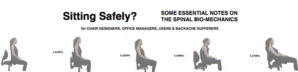
The effort of balancing to maintain this position exercises the small deep (Multifidus) muscles of the back and helps them to regain their reflex supply following an episode of LBP by what is known as proprioceptive neuromuscular feed back. (☛Muscles→) Spinal instability occurs quickly after any spinal disorder and does not easily recover (Hides Wobble ball
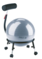
1996) so physiotherapist have developed a wobbly ball for therapeutic exercise when the pelvis is in the correct position. Wilke (1999) gives an intradiscal pressure of MPa 0.5 for
sitting on an ergonomic sitting ball with straight back compared to MPa 0.27 when sitting slouched.     Who wants to sit on a wobbly ball in an office?

The 3M (2T) chair

The 2T concept is simple and requires two STABLE positions. (‘stable’ is used in the ergonomic and not the engineering use of the term) and an unstable intermediate transition mode.  With physiological benefits this can be considered as a mode the 2T then becomes a Triple-Mode (3M) concept.  See 2T CONCEPT a full solution→

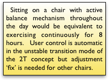
3M. The 2T inherent exercise system.  The concept has a similar effect in the unstable transitional mode with the uncertain advantage that the lateral component, which may strain the facet joints if used continuously, is avoided. So a patient can exercise discreetly and safely following an episode of backache while using a 2T chair.   It also has the advantage that, although no controls are allowed in this mode, it is under the control of the user by entering one of the 2 stable positions at the end of range.

 3M.  Exercise in the transition mode.

  •  Effort, and abdominal muscle activity is required to bring the chair up from the reclining to the upright sitting mode.  The frequency of this maneuver depends on the number of activities performed in this position   As already described, position changes are important for the nutrition of the intervertebral disc which occurs with spinal movement and in particular by the pumping action of the compression/decompression when changing from a supine to upright posture.
  • A further exercise system emerges when the user moves back a few degrees from the forward upright mode and enters the near upright unstable intermediate mode.   Here small amplitude movement is required providing a choice equivalent to  ‘Dynamic Seating’.
  • Additionally 2T chair has to be pulled or swung towards the work-top or pushed away from it, depending on which mode is suitable for the task in hand.
  • Is all-round wobble would preferable to simple anterior/posterior  instability?

3M. Additional systems

These can include a sprung footrest to provide comforting exercise for the calf muscles. These muscles have an important pumping action to aid venous return to the heart and to prevent venous thrombosis.

 3M. A standing mode is easily included in a 3M workstation and would be an advantage in working with a colleague.  This extra mode results in a workstation.   See ☛ 4M concept.→

Stools

A number of bio-mechanically efficient stools have been produced which usually incorporate a FTS, contouring and sometimes iliac support.

Screen Shot 2015-09-30 at 15.15.20
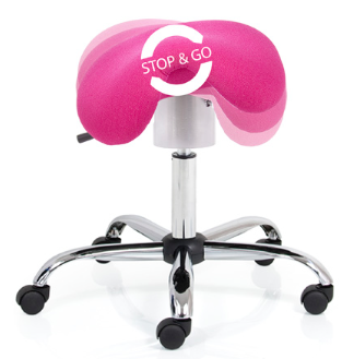
☜ The Pesko ERGO FLEX M 1_1 also incorporates a ‘wobble”.

 Sleep.

A reverse concept!  Short periods of sleep (napping) has benefits resulting in increased productivity   See ☛ SLEEP→

Intervertebral disc angles and the lumbar-sacral junction.

 Spinal configuration of the intervertebral disc angles at the lumbar-sacral junction was developed as a result of hominins adopting an upright (orthograde) stance for efficient bipedalism.   The IV Disc angle is important for preventing pathology and is determined by the overall spinal configurationas as lordosis is protective at the vulnerable lumbar-sacral junction .

The  IV Disc angle and its importance.

The lordotic wedging (IV Disc angle) of the Inter-Vertebral Discs (IVD) have an important function in protecting the discs ((Cyriax JH. 1946 Harrison DD 1998 )  in preventing retropulsion of the disc contents.    This is compromised by some sitting positions.

  •  In moderate extension the position of the IV disc is stable. The  disc contents lie centrally and the back of the joint is closed which prevents their backward displacement. 
  • In flexion the back of the joint is opened up so that the wedge shape is lost and the joint surfaces become more parallel or the wedge is even reversed.  The anterior portion of the annulus fibrosis undergoes compression while the posterior portion is under tensile stress increasing the intra-discal pressure in addition to existing axial compression.  This is an undesirable position as the  altered forces acting on the disc contents, tends to force them posteriorly with a liability for prolapse to occur.
  • The posterior extent of the AF is weaker than the anterior, with fewer lamellae.
  • The effect has already been illustrated with the following diagram.Fl+ax compr
  • The wedge angles of IV discs are the simplest to understand.   It is this angle at the vulnerable L4/5 & L5/S1  ( known as Pre Sacral, PS, 1 & 2 in paleoanthropology) discs that  must be preserved if retropulsion and protrusion are to be avoided.   (Pictures are derived from Francis W. Smith et al.)

Screen Shot 2015-05-31 at 16.32.32

This effect is clearly shown on pMRI scanning.   The wedge angles of IV discs are shown outlined in white on the pMRI scan  (left). (Francis W. Smith et al.)   They are measured more accurately by the shape system used by Meakin et al, shown (right) with dotted  outlines.

Screen Shot 2016-01-31 at 16.04.13In lumbar flexion (left) shows posterior nuclear shift of the nuclear disc contents.  In lordotic extension (right) the nucleus is in the safe mid position.

The Lumbo-sacral (L5/S1) & L4/5 IV disc angles.

The  joints at risk of breakdown are the lower lumbar (L4/5, L5/S1 & to a lesser extent L3/4).   Approximately two-thirds of total lumbar lordosis occurs at the inferior two segments (L4-L5-S1). The total and segmental lumbar lordosis at L4-L5 significantly decreases with age.  As explained these mobile joints are under greater mechanical stress as they adjoin the fused sacral joints which form the posterior wall of the mass of the pelvis.   Having a greater wedge angle than the upper joints, it is likely that this has a protective function which would be lessened if this angle be reduced so that the disc surfaces become parallel or if the wedge is reversed.

The Lumbo-sacral disc angles in sedentary groups liable & not liable to LBP.

Gorman JD reviewing  the x-rays from work by (Pearcy et al.) noted that in the cohort of 11 non- LBP sufferers, a  sedentary group who had not experienced backache in the previous twelve months the standing mean wedge angles were 18° at L5/S1 increasing to 23° with extension.  At L4/5 the mean wedge angle was 16°.

Screen Shot 2013-10-02 at 21.47.22

Note that in this group the wedge angles (Left , in extension) in full flexion (Right)  was preserved although reduced.   Loss of this effect is what is likely to result in joint breakdown, including IV Disc prolapse.   This effect is shown on pMRI scan

Screen Shot 2015-05-31 at 17.33.23

. This study suggests that bipedalism requires a high wedge angle for protection of the lower lumbar discs (IVDs).  Individuals who do not have a tendency for LBP preserve the low lumbar disc wedge angles even when the spine is flexed.  It has been suggested that they may not have used upright chairs when young.   People who are liable to LBP, some 70% of the UK population, do not have this ability to preserve the wedge angle on spinal flexion and the angle may even become 0° or reversed, resulting in retropulsion of the nucleus of the disk towards sensitive structures.

The wedge angles depend on the configuration  of the lower lumbar joints which has been extensively studied and are relevant to chair design.

Vulnerability lumbar angles

Note, in the diagram above, that the upper surface of S1 forms part of both the Sacral horizontal angle and the wedge angle of L5/S1.  The tilt of the pelvis therefore modifies L5/S1 angle.  Upright sitting effects the configuration and reduces the wedge angles.

Lumbar Lordosis (LA)

Lumbar lordosis anglesA new method of calculating  LA from skeletal remains, based on vertebral body wedge angle and the articular process angles, has enabled a study of LA of extinct pre-human and hominin individuals (Been et al., 2010).  Although complicated and with a lack of numbers of extinct forms, it gives a good account of the development of the LA related to bipedalism.

The morphological trend showed the LA jumping from the primitive condition seen in modern apes (22.1°+/- 3.4) to the more human-like LA of early hominoids such as the australopithecines (41° +/_  5)  and hominins such as H. erectus (45°) and then slowly to fossil  H. sapiens (54°8 +/- 14) and modern humans  (51.1° +/-11.0) .  The reduction of the LA in modern humans, at 51.1° from 54°8, who probably sat in chairs since childhood, compared to their extinct Cro-Magnon ancestors, who probably didn’t, might suggest that sitting in chairs has an effect.   The odd ones out are the Neanderthals who show a reversal  with a decreased lordosis (29° +/- 5) when compared with other hominins. with the possible exception of hominins from Sima de los Huesos, who are potentially Neanderthal ancestors. The reduced lordosis of Neanderthals suggests a slightly different gait.  Although having greater body mass and strength, being slower, the reduced LA might imply a reduced LSA or  lumbo-sacral wedge angle with a possibly  increased  liability to LBP and disc protrusion. which might have hastened their demise in the confrontation with H. sap.      The centre of gravity lies further forward later in pregnancy In humans, so that a more pronounced lordosis is required.

 Lumbo-sacral angle (LSA)

More specifically localised to the lumbar-sacral junction which accounts for most of the LA is the Lumbo-sacral angle (LSA) which was used by Abitbol  (Abitbol 1987)  in the study of the phylogenic & ontogenic development of lordosis.

L-sacral angle

The angle is almost nil in non-erect mammals and only slight in monkeys becoming greater in primates which assume an occasional upright stance and bipedal behaviour. It is much greater, about 30˙, in the early hominids.   The angle includes slight wedging of the lower discs  and, to a lesser extent, the vertebral bodies by 5-10°.  This ‘wedge angle’ has an important protective mechanical effect in preventing posterior prolapse of disc components.   In modern humans the LSA is between 70° to  90°.   At birth the angle is about 20° increasing, as the infant assumes the erect posture and  begins to walk, to about 70° at the age of 5 years.

Sacral-horizontal angle

Next consider the Sacral-horizontal angle which indicates the tilt of the pelvis.

Screen Shot 2016-01-31 at 14.50.41

This was used by Pearcy in Xray measurements of a cohort of 11 with no, or low,  incidence of LBP.  This showed a variation between 22-70° and determines the wedge angles of the lower two discs.  The angles being greater than those usually shown in textbooks & journals (Pearcy et al.).  Measurements from this study were further researched by JD Gorman (51).  who, based also on the work of Lovejoy (Tague,  Lovejoy 198652, 53,)  postulated that as the large brained later hominins emerged a larger pelvic birth canal was required.   This was achieved by development of a greater  sacral-horizontal angle which had the effect of increasing the wedge angles of the lowest 2 IV discs and lordosis in additional to the lordosis of early bipedalism.          To see Gorman’s account→

 Paleo-anthropology

Lumbar vulnerability to breakdown at the lumbo-sacral junction originates in the prehistory of the modern and earlier hominins.  Recent changes in human behaviour patterns may have submitted the spine to strains for which it was not developed resulting in increase of the incidence of back pain (LBP) 

The mammalian spine, in it’s various forms, by natural selection has become a beautifully efficient structure.  This has been effected by compromise for often incompatible functions.

  •  The spine has to provide midline central support for the whole body.
  • Give firm anchorage for the weight bearing limb girdles.
  • At the same time it has to allow mobility and respond cybernetically to limb movement and position changes.
  • It has to protect the spinal cord and allow the spinal nerve roots egress.
  • It may also act as a shock absorber to protect the brain.
  • The upright (orthograde) stance adds additional,  sometimes conflicting, requirements.
  • These included partial solution to the lumbar vulnerability at the lumbar-sacral junction of the mobile lumbar vertebrae and the mass of the pelvis and a configuration to limit breakdown.
  • It has resulted in a number of midline spinal curves, backward kyphosis and forward, lordosis and lumbar vulnerability origins may arise from even minor variations.

Orthograde advantages

The adoption of the inherently unstable upright orthograde posture allowed :-

  • Greater height, to see further.
  • Improvement in thermo-regulation required for the savannah environment.
  • Efficient locomotion to escape predators, hunt prey and outrace scavenging rivals.
  • The freeing of the arms from their locomotor function resulted in an increase in manual dexterity allowing new skills and increasing intelligence.
  • Rapid effective response and social organisation were further developed by, and were necessary for, hunting skills.
  • These, in turn, have determined important human social attitudes and behaviour.

Bipedalism began about 7-5 million years ago (MYA) with the advent of the  Hominidae family, which included ancient forms of  modern orangutans, gorillas and chimpanzees and their extinct Photo on 06-12-2013 at 15.17relatives such as Ardipithecus  and  Australopithecus and far down the line, towards modernity, to include humans.  Early hominids were originally arboreal, tree living, creatures that began to walk upright along branches and occasionally on land.  The basic adaption of bipedalism was well advanced in Africa about 3-4 million years ago (MYA). Climate change had replaced the jungle habitat to arid grassland and small shrub Savannah and the Australopethecines were advantaged by  exchanging an arboreal for a  mainly terrestrial lifestyle and development of an upright stance and bipedal gait similar  to that of humans.   The morphological changes are well illustrated by the Australopithecine group, typified by A afarensis and one individual, “Lucy” (or officially Al 288-1), who lived in North East Africa.

Bipedalism and the lumbo-sacral junction  

Anthropoid apes have a straight spine and the torso weight lies anterior to the centre of gravity,   Loading can be brought further back by flexion of the hips and knees, described as   ‘Bent Hips/Bent Knees’  (BHBK, gait)  is required For an upright stance.  This incurs higher energy requirements and a slower gait. Simulation of BHBK walking by humans increases energy consumption by 50%. This is because 80% of energy is conserved by the exchange of potential for kinetic energy by the rising and falling of the centre of gravity.

Bipedalism requires anatomical changes so that the torso can remain balanced upright for most activities and there is an ability to stride forward with the swinging gait which is characteristically human.  This requires the lumbar and cervical spine to be extended in a lordotic configuration so that the axial load of the body is directed down to the ground in a near straight line when standing.   The head, specifically the foramen magnum, is balanced vertically over the plane of the hip joints, in males, and the point of contact of the foot with the ground. This has been achieved by pelvic rotation to enable the hips and knees to straighten.  These lordotic angles and the pelvic rotation are  measurable.

Anatomically, an upright (orthograde) posture and Bipedalism results in :-

  • Lordotic changes to the lumbar spine to avoid walking with bent hips & knees (BHBK).
  • An increase of the IV Disk wedge angle.   This confers a degree of protection from NP retropulsion.
  • Rotation of the pelvic iliac blades for muscles to change from being extensors to abductors ensure pelvic stability.
  • Shortening of the ilium.
  • Relative reduction of the size of the birth canal.

Brain size

The early hominids, such as Homo erectus, had a brain of 900 cc. and its primitive variant, of 1.8 MYA, found at  the Dmanisi (Georgia) site was only 650-780cc. These were probably the earliest hominids outside of Africa (Lordkipanidze, 2005).    H. sapiens, with a volume of about 1300 cc appeared about 130,000 years ago according to the previous ‘out of Africa’ theory’ (Stringer 1970).

Bipedal rats and others 

 Hominids are the only known creatures which are truly bipedal and able to adopt our swinging gait apart possibly wingless birds, such as the ostrich, which are evolved from bipedal dinosaurs. The upright posture occurs in other animals but is usually for short periods and an examination of the skeleton, for example in the penguin, shows a different arrangement with only an analogous appearance of bipedalism.  Performing Japanese monkeys (Macaca fuscata).  can be trained to adopt an upright posture resulting in lumbar lordosis and bipedalism.  Over time some bone remodelling occurs.   However energy expenditure is higher than when plantigrade and they revert to this posture when retired from performing (Nakatsukas 2004).  Slijper, in 1942, gave a detailed account of the changes in the skeleton of a phocomelic goat that had  been born without forelegs. The spine and pelvis had been remodeled with changes suggestive of those found in bipedal bipedal ratanimals.   Rats have been shown (Cassidy 1968) to adopt a bipedal stance and gait if their forelegs are amputated at birth. Their ability to function is remarkable. Their posture and locomotion are surprisingly similar to that of humans and provides the nearest animal model to the human bio-mechanical condition at the lumbar spine. The lumbar spine adapts by becoming lordotic and approximates to that of the human spine and there are changes in the muscles acting around the pelvis. It can be shown that there is increased axial loading on the lumbar spine and a high proportion of these rats develop back disorders which are usually, almost uniquely, only found in humans. These include degenerative changes, disc protrusion, facet joint degeneration and spinal stenosis.

For an associated account  of spinal loading, see ☛ IntraDiscal Pressure→

Next for the obstetric pelvis  and it’s evolutionary importance ☛ see next post → Obstetric pelvis of A afarensis


Positional MRI

 The development of upright positional MRI (pMRI) scanners,  allows imaging of the spine in the load-bearing postures which are relevant to sitting. 

A pMRI study 2006                                                                                                                             The first pMRI study of backward migration of the Nucleus Pulposus (NP) in functional positions was done at the Positional MRI Centre, Woodend Hospital, Aberdeen, U.K by Professor Francis Smith in 2006.   For this study 11 symptom-free volunteers were recruited, aged between 18 and 60 years.

Screen Shot 2016-03-19 at 13.15.20

Results                                                                                                                                            The authors concluded “These results support for the first time the validity of clinical assumptions about disc behavior in functional positions: sitting postures may increase risk of posterior derangement, and prone and supine may be therapeutic for symptoms caused by posterior disc displacement.”   These results confirmed the views of JH Cyriax which he had published as far back as 19452 and subsequently by others3, ,4,5,6.   It also validates the 2Tilt concept as the most effective, possibly the only, way to ensure ‘Safe Sitting’.

Screen Shot 2013-09-25 at 22.33.01The relevant weight bearing positions included sitting upright unsupported, with lumbar support (shown here) and slumped (also shown here). A standing position, which includes lumbar support is also shown here.   Intradiscal pressure in this position was originally measured by Nachemson (1964) showed that intradiscal pressure to be 500-800 N  for a 75 Kg man. See .

Screen Shot 2013-09-25 at 22.32.26Upright sitting, slumped.The usual position and was represented as 30% by students at Cambridge in a quick survey. Intradiscal pressure /load  measured by    Wilke (2001) was 0.48MP  and by Sato  1127 kPa , 800N.  This was less than sitting upright and can be explained by the pressure relieving effect of the abdominal cavity.


Screen Shot 2013-09-25 at 22.31.53

Upright sitting with lumbar (not iliac) support.  As can be seen, the support is applied above the Iliac crest and can be expected to have  an opposite effect at the lower 2 joints (Gorman).   This is shown to be so.


Screen Shot 2013-09-26 at 00.00.37

In the flexed sitting view (left), It can be seen that there is an adverse posterior position of the Nucleus Pulposus (NP) of the vulnerable L4/5 & L5/S1 discs. When compared to non-loaded, uncompressed disc scans (right), the NP has migrated, or in clinical terms there is retropulsion, to the position that is potentially dangerous.

Screen Shot 2013-09-26 at 00.08.35This is precisely what Cyriax had postulated in 1945.


Unexpectedly confirming the view postulated by JD Gorman the scan shows that support at the upper lumbar joints has a reverse, adverse, effect at the important lower joints.   Lumbar v. pelvic support→   See Gorman’s view→

For a review from Working Ergonomics→

pMRI l spt


The pMRI evidence.   Conclusion.

This investigation using Whole-body Positional MRI (pMRI), by FW. Smith, Bashir W (2007) found that the upright position, at 90°, caused disc contents to move the most, while the relaxed position (135°/45° reclined) caused disc contents to move the least. This confirms that the upright position is the worst for the back, while the relaxed position is the best.   From  which the following pictures are derived (arrows, etc, are added).  This confirms the bio-mechanical evidence.

Screen Shot 2016-02-12 at 15.16.18pMRI scan in reclined, relaxed, sitting mode shows the NP in a safe mid-position.  The hip angle is at 135°.    the NP is in the safe  mid-position.   This is practical and preferable and is advocated for the 2Tilt principle in the reclined mode.

Screen Shot 2016-02-12 at 15.18.49pMRI scan in an upright sitting mode shows the NP has translated posteriorly which can culminate in protrusion.   Hips are at an angle of 90° with the seat-pan horizontal.

This is visual confirmation of the bio-mechanical evidence.

SPINAL ANATOMY

Screen Shot 2015-05-31 at 16.12.10Natural selection has resulted in lordotic curves to achieve a bipedal, upright (orthograde) stance in humans.  This has the added advantage of protecting the IV Disc and the posterior spinal structures.   This configuration has to be maintained and is liable to fail with the mid-upright seated posture.    Mechanical breakdown, the cause of common backache (LBP), occurs at two vulnerable spinal levels  

  1. The junction of the mobile neck segments with the mass of the skull.
  2. The lumbar-sacral junction of the lower lumbar vertebrae with the mass of the pelvis.

Lordotic curves, appears on adopting the upright bipedal posture, either phylogenetically as a species or ontogenically as an individual.
spinal anatomy

The foetal spine has a kyphotic C shape  before birth which straightens up by incorporating lordotic changes at the two spinal levels.  It is not straight as in most plantigrade animals although this need not be a biomechanic disadvantage in an orthograde context except would not allow an adequate birth canal.      For further account, see Paleo-anthropology⟶

The Motion Segment.

Motion seg
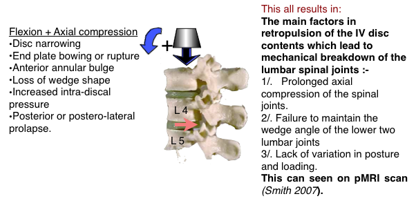
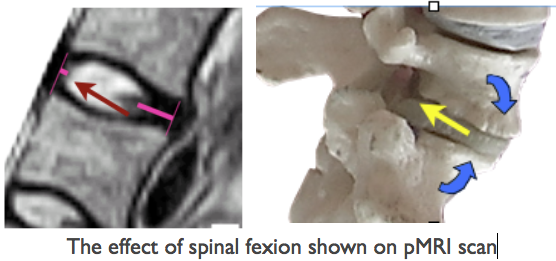
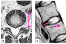
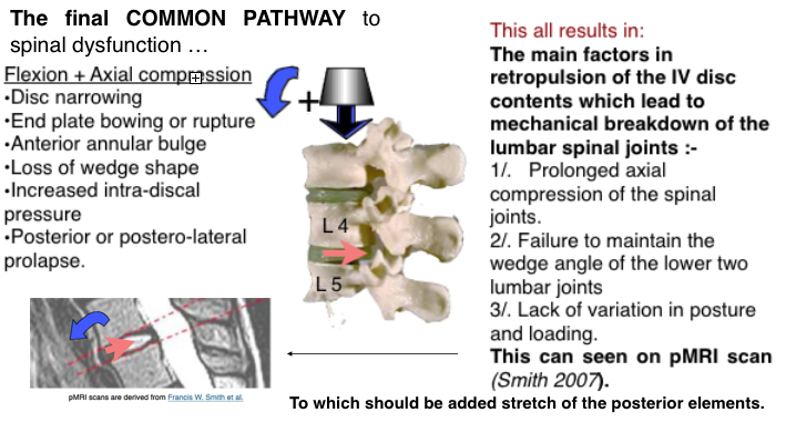
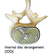
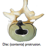
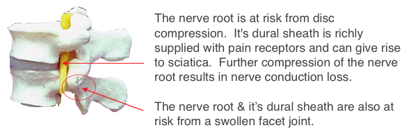
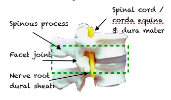
A motion segment consists of the joint, the intervertebral disc (IVD), between the vertebra above and the vertebra below (outlined in green). Included are a pair of associated joints (Zygapophyseal, facet) and various ligaments.  The segment is traversed by a number of important structures (Nerve roots, dura mater, ligaments).   The pathology of structures contained in a motion segment are responsible for symptomatology .   This may vary from backache to sciatica and to a serious equina syndrome.   Breakdown occurs mainly at the lower lumbar joints ( L4/5 & L5/S1) which are highly mobile and at the junction with the immobile mass of the pelvis.   The structures most commonly involved are the posterior spinal ligaments and the inter-vertebral disc (IVD).

The anatomy of the Intervertebral Disc. IV Disc constituents The intervertebral disc  joins two adjacent vertebral bodies allowing the vertebra to move in relation to each other.    Individual joint ranges have been given ( excluding the flexion due to pelvic rotation) as  are L1 8°, L2 10°, L3 13°, L4 16° & L5/S1 17° (Weingarden DS 1991).    It is the sum of ranges at each joint that accounts for the total range of lumbar spinal movement which is very variable depending on age, activities, genetics.                 Next continue and see details ☛ Intervertebral Disc (IVD) →

Spinal Ligaments 

Spinal ligaments


Connect the vertebrae and play an important part in providing the stability of the spine and protection of the disc.A number of midline vertical spinal ligaments lie posteriorly to the lumbar joints.  Their function is to limit excessive flexion, and other, movement  (but vide infra).  They can also give rise to pain and, being visco-elastic, can be permanently lengthened by stretch.     It is possible that ligament stretching in childhood leading to joint instability may be an associated cause of IV Disc degeneration and fundamental to the epidemic of adult spinal breakdown.     See details ☛ Ligaments→ 

Spinal muscles and their stabilising effect.

MusclesWhile ligaments provide passive restraint at the end of joint range, reflex muscle action also provide dynamic support in preserving the integrity  and movers at joints including those of the  spine.        For details of muscle function ☛Muscles

Disc Nutrition & Spinal Movement.

Spinal movement & change of position are important for a number of reasons including IV disc nutrition.       For details see ☛see Disc nutrition

Intervertebral disc angles at the lumbar-sacral junction.

 Spinal configuration of the intervertebral disc angles at the lumbar-sacral junction is important for preventing IV Disc pathology  and developed as a result of hominins adopting an upright (orthograde) stance for efficient bipedalism.  Lordosis at the vulnerable lumbar-sacral junction increased and this also protected the joints.         For details see ☛ Disc (IVD) angles→


Screen Shot 2013-10-13 at 14.47.34

The Intervertebral Disc. (IVD)

The disc is a joint that occupies the ‘space’ that is seen on an X-ray that  lies between two vertebrae. Screen Shot Cyriax

  • Sciatica. Mixter and  Barr in 1934 described the prolapsed intervertebral disc as a cause of sciatica.
  • Backache (LBP).    Cyriax JH in the 1940s Suggested that the lumbar flexion on sitting resulted in  backward translation of the inter-vertebral disc (IVD) contents was a main  element in the cause of backache.
  • To this was added the effect of stretching of the ligaments posterior to the lumbar joints.


The anatomy of the Intervertebral Disc. 

 The intervertebral disc  joins two adjacent vertebral bodies allowing the vertebra to move in relation to each other.    Individual joint ranges have been given ( excluding the flexion due to pelvic rotation) as  are L1 8°, L2 10°, L3 13°, L4 16° & L5/S1 17° (Weingarden DS 1991).    It is the sum of ranges at each joint that accounts for the total range of lumbar spinal movement which is very variable depending on age, activities, genetics.

IV Disc constituentsThe intervertebral disc  joins two adjacent vertebral bodies allowing the vertebra to move in relation to each other.

The Nucleus pulposus (NP):  Occupies 50-60% of the cross sectional area of the disc, is centrally placed, but lies more posteriorly.  It is oval and may have postero-lateral horns and other variations (Jayson MI, 1973).  The nucleus is turgid due to osmotic pressure, maintained by high concentrations of proteoglycans and muco-polysaccharides.   These are present in a semi-liquid hydrophilic lattice gel system.  The intermolecular free water content is approximately 80%  ( Hirsh 1952).  Being contained in the encircling annulus, it exerts horizontal, hydrostatic pressure and being non-compressible effectively resists and redistributes compression forces  (axial loading).  It also acts as a shock absorber.    It is the largest avascular structure in the body and tends to be starved of  metabolic   requirements. These have to be supplied by slow diffusion and are helped by  the “pumping action” of pressure variations due to changes in posture and respiration.

The Annulus fibrosus (AF) is a ring, anchored to the periphery of the vertebral body above and below and contains the periphery of the pulposus. It consists of 15-25 distinct concentric lamellae of collagen fibrils which are  arranged obliquely alternately so that the fibres have an angle of about 50-60˚ to those of the adjacent lamellae. It is especially vulnerable to torsion strains and it may be these that initiate disc degeneration.

Endplates   The Superior and Inferior endplates consist of thin discs of cartilage and are part of the adjacent vertebrae, and not strictly  a part of the disc. They have to be considered as part of the disc mechanics as they act as junctions between the disc and the  adjacent vertebral bodies above and below.    Their blood supply allows nutrients to diffuse into the disc.  Axial compression results in bulging and fracture at a distortion of 8 mm and is the first structure to fail when the disc is not degenerate.

IV Disc detailsAge and degeneration.    

Changes due to aging and those due to degeneration are usually considered together but can be distinguished (Adams A, Bogduk  Adams, Michael).  Aging changes in the disc, both biochemical and biomechanical, start in infancy and are progressive (Kraemer 1995). By the age of 50 disc degeneration is universally  present (Andersson  1997).   The morbidity curve for discogenic symptoms does not follow the curve for degeneration.  The commonest period for symptomatic disc protrusion is the 4th and 5th decades although the process may have started much earlier.

By middle age osmolarity still exists in the nucleus although hydration and proteoglycans are reduced.  With degeneration, the stress profile alters with variable and irregular peaks (Adams, Bogduk 2002) and axial loading results in stress peaks particularly in the posterior annulus.  This is the area where the disc becomes more vulnerable to breakdown and symptoms are likely to occur.

A degenerate disc is one with structural failure combined with accelerated or advanced signs of aging.  This is hastened by

  • Damage to the endplates or annulus, which  can occur with  excessive Intra Discal Pressure (IDP) or strains, particularly if coupled with bending.   
  • Instability of the motion segment  occurs with laxity of the controlling ligaments and atrophy of the deep controlling spinal musculature, the Multifidus muscle complex, which atrophies following joint injury (Hides 1996).
  • As ageing (Type II) progresses to degeneration (Type III), the annulus becomes increasingly

Screen Shot 2013-11-19 at 21.31.38fissured and weakened. A variety of disruptions of the fibres of the annulus are described including radial clefts and peripheral ‘rim’ lesions which are parallel and near the circumference of the disc (Schmorl 1971) and   are easily detected, markers of impaired disc function.

These, and fissures in the endplates due to excessive compressive forces hastens further  nuclear degeneration and are likely to lead to nucleus pulposus prolapse. (Vernon-Roberts 1975).

  • Cyriax emphasised that radiological evidence of OA, in the absence of relevant physical signs, was irrelevant to clinical diagnosis  (Cyriax JH. 1945).
  • Excessive spinal loading augmented by spinal flexion occurs on upright sitting.
  • Immobility results in reduced nutrition of the disc.

Screen Shot 2013-11-19 at 21.29.50Spinal breakdown may ultimately occur with excessive biomechanical trauma which can be described in engineering terms – compression; torque; sheering; bending moments; distraction.  Motion segments where the disc was intact failed, in vitro, at an average torque of 881.4×10⁶ dyne-centimetres (Farfan 1970).  Although it has been shown that an intact intervertebral disc can withstand a considerable direct compressive force, this changes in the commonly degenerate disc, when combined with flexion. The nucleus is then liable to protrusion, prolapse and extrusion (Adams et al.,2000b; Natarajan et al., 2004).

In old age the lumbar lordotic curve, which in early childhood is secondary to the need to stand upright, eventually reverts back towards the primary prenatal curve into flexion.  The disc wedge shape is lost and there is osteoporotic wedging of the vertebral bodies.  Disc prolapse in the elderly is rare as the disc tends to be contained by osteophytes and the disc contents are replaced by fibrous tissue resulting in fibrous ankylosis.  At this stage the  main pathology is due to osteoporosis or spinal stenosis, which is not common.

See Disc Nutrition and movement  →

Effect of the upright sitting mode on IVD pathology

  • The conventional mid upright sitting posture causes backward tilting of the pelvis resulting in reduction, or even reversal, of the protective disc wedge angles at the lowest 2 lumbar joints (L4/5 & L5/S1) where mechanical spinal breakdown is commonest.
  • Posterior disc protrusion was put forward originally by Cyriax (1945) as the main cause of backache.  He also emphasised the importance of the disc wedge angle in preventing prolapse with the adage “Preserve the lumbar lordosis”.      For details see ☛ Disc (IVD) angles→
  • Gorman showed that This effect is augmented by lumbar support directed to above the L4 vertebra  .  The predicted effect is retropulsion of the disc contents (NP) and is confirmed by pMRI studies (Smith, 2006).
  • •Pelvic tilting also stretches of the lumbo-sacral and supraspinous ligaments which can become irreversible and result in pain and joint instability leading to CTD and earlier degeneration (see Anatomy/Ligaments→).

Clinical effects

   If the annulus is unable to contain the increased hydrostatic pressure of the nucleus, while being stressed vertically and simultaneously stretched posteriorly by forward pelvic tilting, retropulsion of the nucleus pulposus becomes more likely.   Anterior bulging also occurs and in time becomes contained by osteophytic (‘Parrot’s beaks’ in French) outgrowths from the adjoining vertebral margins.   These are evident on x-rays and are silent clinically.  This is in contradistinction to disc bulging with a backward or a postero-lateral protrusion where impingement is likely on a number of pain sensitive structures.   Herniation or rupture of the annulus can then occur  with posterior nuclear or, more commonly, a postero-lateral protrusion which can threaten the emerging nerve root.  A large midline extrusion can rupture the posterior longitudinal ligament (PLL) resulting in the surgical emergency of a Corda Equina syndrome

Cyriax and the IV disc.  An historical diversion.

Discussion of IV discs and LBP without mentioning Cyriax is equivalent to discussing Natural Selection without mentioning Darwin.   Mixter & Barr in the USA having identified IVD prolapse as the main cause of sciatica, Cyriax, at St Thomas’, London, postulated it as the main cause of backache (LBP)  (Cyriax JH. 1945).  At that time the IVD was regarded as aneural and avascular.   I can actually remember a lecture by Prof HA Harris to this effect at Cambridge. so Cyriax had to postulate it’s prolapse and impingement on various adjacent structures as the cause of symptomatology.   These included the Posterior Longitudinal Ligament (PLL), the anterior surface of the Dura Mater for ‘extra-segmental’ pain and the dural sheath of the emerging spinal nerves for sciatica, culminating in nerve root conduction loss or, rarely, a cauda equina syndrome.  He used a 50 ml. 1/2% Procaine by caudal epidural for confirmation.   On various grounds he excluded facet joint, ligaments and the sacro-iliac (SI) joints as causes which degraded his development of a systematic clinical functional examination of the spine but not of peripheral structures.  This not only enabled an accurate localisation of soft issue lesions but also  diagnosed ‘psycho-genic’ (‘psycho-social’, pg) as a common cause or overlay and that in these cases physical treatment should be avoided.  Recently this has come to be considered by some as the main cause of LBP.

Screen Shot 2016-03-18 at 11.06.39

Screen shot 2013-10-30 at 17.13.39“Lordosis…..directing it (pressure) away from the sensitive structures.”   (that lie posterior to the joint, the Ligaments, nerve root, dura mater).

From,  Cyriax JH. Illustrated manual of Orthopaedic Medicine. 1982. p. 140

At that time some of Cyriax’s contemporaries criticised his hypothesis as ‘unscientific’ as it was based mainly on observation and not on his own controlled trials.    Subsequently his views have become mainstream and used as a basis for treatment by a large body of therapists, medical and physiotherapeutic, worldwide.  Later research showed pain receptors (nociceptors) in the posterior part of the annulus (AF) and the surrounding structures and so could be reconsidered as causes of LBP.    This simplified the Cyriax hypothesis as direct impingement was no longer a necessity to explain the varied symptoms. Stretching of the posterior ligaments has also come to be recognised as a nociceptive and etiological factor (See Ligaments/Cumulative trauma disorder→).   Screen Shot 2019-01-04 at 13.43.15

The Cyriax archive devolved  to me and I gave it to the  ☞Welcome institute  for safe-keeping.  It included his entire patients notes which are a valuable research resource as they were detailed and systematic.

To continue see detailed sites on the biomechanics factors affecting IVDs.   Next see ☛ IV Disc angles→ and the lumbar-sacral junction.

Spinal Ligaments and CTD.

Spinal Ligaments connect the vertebrae and play an important part in providing the stability of the spine and protection of the disc.   Adams et al. (1980) showed that the supraspinous-interspinous ligaments segments are the first ligamentous tissues to become stressed with forward bending of the lumbar spine.

Ligament function related to the spinal ligaments

  • The function of ligaments is to restrict movement of a joint in a specific direction. Laxity through rupture or lengthening, can allow joint movement beyond the capacity of the muscles to act normally and also allows reduction of the protective wedge angle of the lower lumbar joints.
  • Ligaments also have a proprioceptive function allowing the CNS to determine position and feedback for correction.
  • Contain nociceptors and are a source of pain.

  A number of midline vertical spinal ligaments lie posteriorly to the lumbar joints.  Their function is to limit excessive flexion, and other, movement  (but vide infra).  They can also give rise to pain and, being visco-elastic, can be permanently lengthened by stretch.

Spinal ligamentsIt is possible that ligament stretching in childhood leading to joint instability may be an associated cause of IV Disc degeneration and fundamental to the epidemic of adult spinal breakdown.

The Interspinous ligament is  a thin septum between borders of each adjacent spinous process. Fibres  fan out in an antero-caudal direction at an angle of about 20˚ to the horizontal32. Tension imposes an anterior shear force on the disc.  This arrangement may also ‘even out’ the flexion ranges at each of the spinal joints (Gorman. 1987) .  Stretching  plays a part in the inhibition of the multifidus muscles (Solomonov 1999).

Supraspinous ligaments are formed from the midline element of the lumbo-dorsal fascia (Thoraco-lumbar fascia, TLF).  Strong and cord like, they connect the tips of each spinous process becoming taut at extreme of, and limit, lumbar flexion.        They  are the first structure to fail, at extreme flexion (Rissanen PM 1960) (Adams, Hutton 1980) and are richly supplied with nociceptive (pain)  small nerve fibres.  However, they don’t extend beyond L4 to stabilise the important lumbo-sacral junction and are replaced by complex arrangements  of bands of collagen fibres derived from the deep layer of the thoraco-lumbar fascia (Bogduk).  The one  from the tip of the L4 & L5 spinous processes to the posterior superior iliac spine and beyond,the ilio-lumbar ligament, has the greatest offset and so is likely to be the most important in limiting flexion range (Adams, Bogduk 2002).   (NOTE. In my clinical experience this structure was commonly involved in chronic LBP. HAS)

Screen Shot 2013-11-04 at 13.56.25The likely forces that must be resisted by the ilio-lumbar and the supraspinous ligament (shown in blue) when sitting in the usual lumbar support seat, which allows backward tilting of the pelvis, with a bodyweight 40kg (excluding the legs) can be calculated.  Simple moments about the centre of the L5 disk suggests a ligament tension of about 70 kg (700 Newtons).  This is probably a worst-case estimate.   (JD Gorman)

In the early days, working at St Thomas’s, at that time being the low man on the totem pole, it fell to my lot to infiltrate the supraspinous ligaments with a sclerosant solution as a stabilising procedure.  I soon noticed that this could result in unexpected immediate and complete symptomatic relief, the patient walking away rejoicing.  I could identify the indications where this was likely and could postulate that the ligamento-periosteal attachment was the site of the lesion.  This did not go down well with Cyriax who was of the opinion that pain did not originate in spinal ligaments.  I asked him why if pain could arise in any other ligament, “Why not spinal ligaments?”.   “Yes , very odd, but it is so.”    I also noticed that I could identify the top end of the L5/S1 (lumbo-sacral) ligament, a common pain source, but the bottom (sacral) end seemed to have got lost.  This was explained by Bogduk’s account of the anatomy showing that this arrangement is modified at the important L4/5 and L5/S1 levels and, as mentioned,  flat ligaments fan out from these spinous processes to be attached to the iliac crest and sacrum.  providing a broader and stronger caudal attachment (Bogduk 1984) (Heylings 1978).

The Ligamentum flavum  is a strong elastic yellow ligament which connects the adjacent borders of the laminae of the posterior vertebral arch, and with them forms the posterior wall of the spinal canal. It protects the neural elements in the canal (Ramsay 1966).   The ligament may become thickened with degenerative changes and lose its elasticity which may result in kinking which can encroach on the spinal canal  resulting in narrowing (spinal stenosis described by Verbiest).  Doubt  has been expressed as to whether this occurs but I must admit to having myself often seen thickening and kinking at surgery.

Posterior longitudinal ligament. This is important strong and ribbon-like extending the whole length of the spine, forming the anterior wall of the spinal canal.     It is attached to the vertebral bodies and spans the intervertebral discs.   This topography limits posterior protrusion of the disc contents and being weaker or absent laterally, protrusions tend to be directed  postero-laterally where the emerging nerve roots, in their dural sleeve, emerge.  Complete rupture of the ligament allows a dangerous mid-line extrusion of disc contents which threatens the adjacent spinal corda equina (the spinal cord does not extend to the lower lumbar region) in the spinal canal. This can result in nerve root conduction loss and paralysis of the legs and sphincter loss which can be permanent if not relieved by emergency surgery.

Capsule of the facet  (zygapophyseal)  joints

The facet joints are bilateral accessory joints between the articular processes of adjoining vertebral arches    They guide and limit movement provide 39% of the intervertebral joints’ resistance to bending (Adams 1980) and also account for some of the axial loading on the lower part of the spine, especially in excessive lordosis with the transfer of the axial loading from the nucleus (NP) onto the facet joints (Adams 1980Adams 1981) which can also give rise to pain (Kuslich 1991).  This increases with disc degeneration.  The capsular ligaments can become stretched and the joint can become degenerate and disorganised.   I found the posterior capsule of these joints to be clinically identifiable and easily and effectively treated although trials of transcutaneous electronic nerve stimulation, and facet joint blocks have not reached significance (Jackson RP 1992).

Thoraco-lumbar fascia (TLF, lumbo-sacral fascia)  Simplistically, the TLF acts as an extensor and anti-flexor of the lower lumbar spine (L2-S1).    Consisting of a trilaminar investing fibrous membrane which encloses the trunk ( like a body-stocking) some fibres become condensed to form ligaments and tough tendinous aponeuroses.      The detailed anatomy is complicated.   The superficial layer of the TLF  posterior lamina, through the aponeurosis, gives spinal attachments for the latissimus dorsi muscle and, through the lateral raphe, for the internal oblique abdominal muscle which also has a more direct attachment through the deep layer (of the TLF posterior lamina) to the tips of the spinous processes.   (See below).     The view that ligaments are merely passive restraints on excessive joint movement has to be modified for the spine.  As shown above the TLF mesodermal structures consisting of fascia, aponeurosis, ligament and tendon anatomically blend into each other and subserve similar functions with great complexity.      Intra-abdominal pressure also tenses the TLF (Gracevetsky 1985) and supports a  proportion of the body weight which may be protective in slumped ethnically determined ‘natural’ sitting postures.

Cumulative Trauma Disorder (CTD)

 Spinal ligament lengthening, which can start in early life, may be postulated as the basic faulty link in the chain that leads to muscle dysfunction, disc degeneration and internal disc disruption (IDD).

In adulthood the stretching of posterior spinal ligaments that occurs during a work period when sitting in spinal flexion does not fully recover before the next session.  This may have a cumulative effect over a day and even over many days, weeks, months and years becoming chronic.   Solomonow, using an in vivo feline model, showed that creep developed in the viscoelastic tissues of the spine, over a series of only three equally long of such static flexion/rest sessions, under a moderate physiological load, did not fully recover during an equally long rest period and accumulated to a value of 52.77%.  Seven hours of rest were not sufficient to allow full recovery of the creep developed during 30 minutes cumulative work, leaving residual creep for the ‘‘next work day’’ (Solomonow et al 2003). Sitting postures, which allow prolonged stretching of the ligaments may lead to permanent lengthening and instability,  an important factor in disc degeneration  (see also, Ischial off-load). It  is particularly relevant to poor posture in childhood (See post, School chairs & desks→). 

A 2Tilt (2T) chair in the reclined mode, the the spine is fully and correctly supported , for prolonged work, particularly at the lumbo-sacral junction.   See  2T CONCEPT a full solution→.     This may be an uniquely important factor in the avoidance of CTD.

Nerve supply of the IV Disc.

 We have come a long way since, as a medical student, I was taught that the nerve supply of the intervertebral disc was non existant .

Cyriax, working at St Thomas’, London, a pioneer in identifying the IV disc as a main cause of Low Back Pain (LBP).   At that time, in the late !940s, IV Discs were thought to have no nerve supply and so he had to postulate that the symptomatology of IVD pathology was determined by the structure against which an IVD herniation impinged. Subsequent research found pain receptors via an extensive distribution of small nerve fibres and free nerve endings in the superficial annulus (AF) of the disc which simplified this view.   He also postulated that if a structure was impinged upon or stretched its nociceptors would be activated which has also been confirmed by pain-provocation studies  and modulation by psycho-social aspects confirmed (Kuslich 1980)..      In addition mediation by biochemical agents has been identified.

Nerve root pain (sciatica)

Pain is in the distribution of the individual nerve root (see right→).  Conduction loss results in motor , sensory and reflex loss. Onset usual follows episodes of LBP but may be of initial occurrence and the presenting symptom.  This was termed ‘Primary postero-lateral protrusion’ by Cyriax.  A large posterior protrusion could cause rupture of the Posterior Longitudinal Ligament (PPL) resulting in a cauda equina syndrome.  A surgical emergency.

Screen Shot 2015-06-02 at 19.45.15

A better diagram can be seen at precisionhealth →

Other causes of LBP

In the case of facet joint pain, several mechanisms were identified including small nerve fibers and endings (Yoshizawa 1980).

The sacro-iliac joint has also been shown to be a significant, yet poorly understood, source of low back pain (Schwarzer , Bogduk 1995).  These studies show that there is a logical and well demonstrated rationale to expect that mechanical stimulation of these spinal structures can lead to low back pain perception and reporting.

Moderate pressure on the dorsal root ganglia resulted in vigorous and long-lasting excitatory discharges that would explain sciatica. In addition, sciatica could be explained by excitation of dorsal root fibers when the ganglia were exposed to nucleus pulposus material.

Free nerve endings have been confirmed in the Posterior Longitudinal Ligament (PLL), which Cyriax claimed as the main origin of LBP, by Bogduk, (1991), (Cavanaugh, Kallakuri, and Ozaktay1996;) (Blagoev, 1998) and small fibers and free nerve endings in the nerve root sheath.   Earlier, in 1945 Cyriax showed that these various pain sources could be blocked by weak local anaesthetic (Procaine ½%) by caudal epidural. In the case of radicular (sciatic) pain this relief was often permanent (Cyriax 1945).   Later Bupavicain and steroid were used.   This procedure, whether by the caudal or trans-lumbar route, gradually became mainstream and many patients were spared the potential complications of surgery.

In addition to the usual nociceptive function neuromuscular control is regarded as important to prevent spinal motion segments from buckling in shear, axial rotation and/or bending (Crisco and Panjabi, 1992; Adams, 1995).

Functional clinical examination can usually identify an anatomical localised organic lesion as the source of the pain, and determine whether complicated by, or due to, psychogenic overlay.  The psycho-social input has become more recognised and studied in the last 20 years.   The terms ‘Non-specific’ or ‘Postural’ Backache are best avoided.    Cyriax famously remarked “I know of no anatomical structure called the ‘Posture’.

Next see  ☛Disc nutrition & movement→

Allied & detailed sites

Spinal muscles and their stabilising effect.

While ligaments provide passive restraint at the end of joint range, reflex muscle action also provide dynamic support in preserving the integrity  and movers at joints including those of the  spine.  

Function.

Co-contraction of muscles preserve the upright posture of the trunk and allow the spinal joints to be stiffened, stabilized, and moved in a well-controlled manner.  The antagonistic force, and secondly by the additional agonist force required to balance this antagonistic force, together impose greater loads on  tissues of the musculoskeletal system including the IV discs.  This occurs in work activities which involves handling heavy loads, and also includes the flexion that occurs when sitting upright and unsupported on an horizontal surface.

Lumbar extensor muscles. 

MusclesThe main movers of the lumbar spine are the most posterior group of muscles.

   They control flexion and exert extension, having extensive origins from the transverse processes the lumbar vertebrae and with adjoining rib surfaces of the thoracic vertebrae, consisting of the :-The arrangement is complex with decussating fibres across the midline.   The fibres form fascicles which have a widespread attachment to the ilium, including an extensive attachment via the extensor aponeurosis, and to the posterior surface of the sacrum.    The most relevant to the control of spinal flexion at the lumbo-pelvic junction are those that extend from the thorax to the sacrum and the posterior iliac crest as these have the greatest offset and mechanical advantage. The parts of the muscles that arise from the thorax form the  Erector spinae aponeurosis, whose medial fibre form the Longissimus Thoracis attach to the posterior surface of the sacrum (Bogduk 1992 1997).   The flat abdominal muscles, the transversus abdominis and external oblique muscles, have an attachment to the lateral raphe of the  extensor aponeurosis and thus to the erector spinae.   The latter also attaches to the iliac crest. They are, in part, responsible for maintaining abdominal pressure which has a spinal stabilising effect (Gracevetsky 1985) but may increase axial pressure (Granata 2000).

The over centred position.

Screen Shot 2013-09-17 at 01.24.31If, on lumbar flexion, the wedge angle is lost then these attachments now lie below the lower lumbar joints. In engineering terms this an over-centre mechanism.  The ability of the spine to resist flexion forces will reduce to almost nothing beyond a certain angle of flexion of the lowest joints. (Text & diagrams after Gorman JD with permission).   Note that approximately two-thirds of total lumbar lordosis occurs at the inferior two segments, the L4-L5 & L5-S1 (Kamali 2003).

Muscle atrophy (wasting)

Reflex muscle wasting is a usual accompaniment of any spinal joint disorder and this has important implications.  Following an acute episode of LBP, wasting has been shown (Hides, Stokes 1994), by ultrasonography, to occur surprisingly quickly, usually within 2 weeks.    The wasting is confined to the specific spinal level of the lesion and on the side of the lesion and involves mainly the multifidus muscles at a local level.    These changes result in weakness, fatiguability and loss of coordination and are likely to be a factor in the perpetuation and recurrent nature of chronic LBP (Hides, Richardson 1996).  The changes in the muscle fibres are complex and their cause not completely understood.  Reflex neural activity may play a part.  This has been found to be the case in analogous conditions of the knee (Johansson 1991)  and ankle for which treatment using an unstable standing platform (‘wobble board’) for balance has been found effective.    These atrophic changes are not reversed spontaneously even after return to normal activity.  It has been  shown that specific stabilising exercises for the multifidus  help recovery but these are difficult and specialised.    Stabilisation is an important factor in preventing chronicity and LBP recurrence (Saal & Saal 198941, Wilke 199571).  It will be shown later how the unstable mid position of the 2Tilt chair helps in this rehabilitation.  The addition of some side-to-side instability might be advantagous.   A number of stools and chairs with unstable, seats, or wobble balls, have been developed.

 Studies by Sweetman at Swansea suggest that heavy work provokes a facet joint syndrome but is protective for the disc.   In previous studies the various LBP syndromes were not distinguished and so statistical results tended to be inconclusive.   This would confirm the observation that people engaged continuously in heavy work, or other exercise, having strengthened their back musculature, have a decreased liability to back ache.     This is in contradistinction to the suburban weekend gardener who attends my spinal clinic on a Tuesday morning with a facet joint sprain.   Unlike excessive strains, usual activity is not a significant aetiological factor in disc breakdown although may exacerbate minor backache.  Many patients attend saying “I think I’ve pulled a muscle in my back” or even “My doctor tells me that I’ve sprained a muscle in my back”.      Clinically this is a rare occurrence and muscles are usually only damaged by trauma in rough contact sports and even then a sudden over-strong muscular contraction snaps a bony lever at its attachment.   This observation is easily tested by exerting a strong resisted spinal extension, so that the spinal extensor muscles are isometrically contracted without any joint movement.  Pain or weakness on this action indicates a muscle lesion and is so rare that it is omitted from the routine examination by most orthopaedic physicians.   The role of pain arising from muscles is uncertain and controversial. Tender knots and nodules which are commonly felt in the muscles and can command much attention, which in the past were known as ‘fibrositis’, are usually due to localised spasm which is a referred phenomenon from a lesion in a deeper structure.

Muscles moves the spinal joints which alters intra-disc pressure which results in the pumping effect, responsible for disc nutrition ⟶

Next see Disc nutrition→

Allied & detailed sites

Loading

There is some confusion on the extent and role of spinal compression.

2 worst combinations

Axial loading & intra-discal pressure (IDP, IAP).

Early in vitro loading studies (Jayson, 1975) showed little or no effect on intact discs in the young.  In most cases, failure occurred in the vertebral endplate rather than by nuclear (NP) prolapse.  Cadaveric discs from males between the ages of 22 and 46 could, on average, withstand single loads of over 10,000 N before failure occurred (Adams AC and Hutton 1982).     When hyper flexed in vitro discs prolapsed at an average of only 5,400 N instead of 10,000 N  (Adam et al.2002).

Rather than through peripheral nuclear prolapse, in most cases, the failure was in the vertebral endplate, resulting in Schmorl’s nodes.   Since the disc is a largely avascular structure, the health of the endplate is critical for nutrient exchange, and even small failures may hasten the degenerative process.

In vivo studies


Loading studies, in vivo by direct measurement, in addition to degeneration and joint flexion,  is complicated and fraught with variations in the state of the disc, neuromuscular activity, facet joint load, ligamentous laxity or  tension.  However all show Intra Discal Pressure (IDP) to be lower in the reclined posture than with mid-upright sitting. IDP studies were pioneered  by Nachemson  (Nachemson A. 1964) who   showed that intradiscal pressure (IDP) to be 500-800 N  for a 75 Kg man, while standing,  Sitting  IDP was higher.

  • Screen Shot 2015-06-09 at 16.57.21
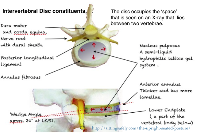
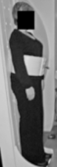
Standing  100  ( In standard units based on %.)
  • Lying supine  (face upwards)  25
  • Forward flexion (bending forwards) 150
  • Sitting in flexion 185,     sitting and lifting     280,
  • Lifting  220,
  • Cough   140,   Laugh  150
  •  Later modified to lying, 150-250 N,      sitting 700-1000 N.

Later work by others contradicted this and reported near identical pressures for both sitting or standing, being approximately 0.30 MPa (Schultz , 1982).

IVD pressures


Modifying factors occur with :-

  •  Degree of disc degeneration (Sato et al., 1999)., which is eventually nearly universal.
  • Joint flexion (Sato et al., 1999).which occurs with unmodified upright sitting.
  • prolapsed discs occurred more frequently when the vertebral segments were wedged to simulate extreme forward bending of the spine (Adams and Hutton, 1982). In this position, the anterior portion of the annulus fibrosis undergoes compression while the posterior portion is under tensile stress. Over 40 percent of the cadaver discs tested by Adams and Hutton (1982) prolapsed when tested in this hyperflex posture, and with an average of only 5,400 N of compression force applied. This finding shows that the disc is particularly susceptible to bending stresses.
  • In a later study in which Adams and Hutton (1985) simulated repetitive loading of the disc, previously healthy discs failed at 3,800 N, mostly through trabecular fractures of the vertebral bodies.
  • Taken together, these studies indicate that the disc, especially the vertebral endplate, is susceptible to damage when loading is repetitive or when exposed to large compressive forces while in a severely flexed posture.
  • When combined with flexion IDP loading can produce regions of higher stress in the posterior annulus strongly associated with disc prolapse (McNally et al., 1993).
  • The state of the disc,  it’s volume, surface area,  previous activity and hydration.
  • In this respect it should be noted that during 7 hours of a night’s sleep,  intradiscal pressure may change as much as from 0.1 MPa to an average of 0.24 MPa in the morning, an increase of over 100% (Wilke 1995) (Zander T, 2010).
  •  The zygapohyseal (facet) joint function and pathology becomes more pronounced with the disc narrowing that occurs with OA..
  • Reduction of IDP occurs with extreme spinal extension (lordosis) when the neural arches become compressed together (Adams 1994).   This transfers load to posterior elements, the posterior extent of the annulus, ligaments and facet joints.   These are supplied with nociceptors (Kuslich 1991).
  • Compression (IDP) is not simply due to the weight of the body that lies above the level of the specific disc.  The addition is due to co-contraction of the muscles that preserve the upright posture  of the trunk and allow the spinal joints to be stiffened, stabilized, and moved in a well-controlled manner.   This increases in work activities, which includes the position that occurs when sitting upright, and impose greater loads on the discs and other tissues of the musculo-skeletal system (Wilke & Wolf 1995)  and in some spinal flexion deformities.
  • Relevant to disc protrusion are torsion and shear forces when applied to a vulnerable disc.

Considerations on the later studies on intradiscal pressures.

Studies, from Japan, Sato  (Sato et al., 1999), using a piezoresistive sensor in the transducer needle detecting the pressure laterally at a window and not at the needle tip, confirmed the pioneering findings of Nachemson and examining the changes in the more relevant L4/5 disc rather than the L3/4.

  • This later work showed the load of the spine changes in a curvilinear fashion, when moved from a flexed to an extended position.  In healthy subjects with an average body weight of 73 kg and an average L4–L5 disc cross- sectional area of 16 cm2 the compression pressure for prone lying was found to be 144 N, and for Standing  800 N & Sitting upright  996 N, confirming Nachemson’s findings.  Also  intradiscal pressure fluctuate with breathing, which may help disc nutrition when sitting in a cramped position.  Reduction in hydration which occurs with degeneration of the  N. Pulposus significantly reduced the pressure as degeneration progressed.    Sato also radiographically evaluated the disc wedge angle for each measurement and concluded that the spinal load was highly dependent on this angulation.

More recent works differ from that of Nachemson and Sato in that pressure on sitting upright is similar or only slightly more than when standing.  As subjects without LBP or radiological evidence of degeneration were used, this has little relevance to the effect of IDP on discogenic effects.

  • Wilke (Wilke et al. 1999) in a study on a single subject using telemetry to record from an implanted transducer  transmitting data, during a variety of daily activities, over 24 hours.  Recorded pressures included :-
  1. Standing intradiscal pressure of  0.48 MPa,
  2. Relaxed erect sitting without, or not using the  backrest, slightly slumped, as is usual was 0.44 MPa.
  3. With lumbar support 0.33 MPa Sitting with actively straightened back with no support was 0.55 MPa.
  4. Later work (Wilke et al. 2003) showed loading to be thee times higher during unsupported flexion and almost three times greater than with relaxed sitting.

Disc protrusion. Pathogenesis

To assess the biomechanical factors involved in the protection of the vulnerable lumbo-sacral junction, knowledge is required of :-

  1. Intradiscal compression (IDP). The intra-discal measurements are those of the NP which being aneural is not a pain source..
  2. Disc wedge angle of the disc (Sato 1999).  Extension transfers load to posterior elements, the posterior extent of the annulus, ligaments and facet joints which have nociceptors (Kuslich 1991).
  3. Position of disc contents (NP)  as shown by pMRI (Smith 2006).

Other views on discogenic pathogenesis           

Garun Garg at NIOSH  concluded that intradiscal pressure alone  as a risk factor is minimal at best. Protrusion can occur at low IDPs (Kumar, S. (1998, 2001).   The subject was reviewed in Australia (Clause Hides 2008), and concluded “If sitting is a greater threat for development of low back pain than standing, the mechanism is unlikely to be raised IDP.”  but mainly quoting sources confined to studies on intact discs.

Next ☛Intervertebral disc angles and the lumbar-sacral junction.

Lumbar vulnerability and Paleoanthropology.


Lumbar vulnerability to breakdown at the lumbo-sacral junction originates in the prehistory of the modern and earlier hominins.  Recent changes in human behaviour patterns may have submitted the spine to strains for which it was not developed resulting in increase of the incidence of back pain (LBP)
 

Screen Shot 2013-10-23 at 13.57.25
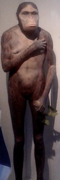
Development by natural selection has resulted in the mammalian spine, in it’s various forms, becoming a beautifully efficient structure.  This has been effected by compromise for often incompatible functions.  Lumbar vulnerability origins may arise from even minor variations.

Vertebrate spinal functions

  • The spine has to provide midline central support for the whole body.
  • Give firm anchorage for the weight bearing limb girdles.
  • At the same time it has to allow mobility and respond cybernetically to limb movement and position changes.
  • It has to protect the spinal cord and allow the spinal nerve roots egress.
  • It may also act as a shock absorber to protect the brain.
  • The upright (orthograde) stance adds additional,  sometimes conflicting, requirements.
  • These included partial solution to the lumbar vulnerability at the lumbar-sacral junction of the mobile lumbar vertebrae and the mass of the pelvis and a configuration to limit breakdown.
  • It has resulted in a number of midline spinal curves, backward kyphosis and forward, lordosis.

Orthograde advantages

The adoption of the inherently unstable upright orthograde posture allowed :-

  • Greater height, to see further.
  • Improvement in thermo-regulation required for the savannah environment.
  • Efficient locomotion to escape predators, hunt prey and outrace scavenging rivals.
  • The freeing of the arms from their locomotor function resulted in an increase in manual dexterity allowing new skills and increasing intelligence.
  • Rapid effective response and social organisation were further developed by, and were necessary for, hunting skills.
  • These, in turn, have determined important human social attitudes and behaviour.

The development of Agriculture occurred in the Middle East only about 10,000 years ago, probably as a result of climatic changes (Rowley-Conway, 2007) and allowed a more settled lifestyle and may have involved more non-erect, sedentary activities, such as food preparation and repetitive use of groundstone tools for grinding grain.  This was mainly a female’s activity while the men were away happily hunting.  Archeological evidence shows an increase of spinal degeneration at this time which affected females more than males (Molleson 1994).

Spinal curvature.   DEVELOPMENT

Bipedalism began about 7-5 million years ago (MYA) with the advent of the  Hominidae family, which included ancient forms of  modern orangutans, gorillas and chimpanzees and their extinct relatives, Photo on 06-12-2013 at 15.17such as Ardipithecus  and  Australopithecus and far down the line, towards modernity, to include humans.  Early hominids were originally arboreal, tree living, creatures that began to walk upright along branches and occasionally on land.  The basic adaption of bipedalism was well advanced in Africa about 3-4 million years ago (MYA). Climate change had replaced the jungle habitat to arid grassland and small shrub Savannah and the Australopethecines were advantaged by  exchanging an arboreal for a  mainly terrestrial lifestyle and development of an upright stance and bipedal gait similar  to that of humans.   The morphological changes are well illustrated by the Australopithecine group, typified by A afarensis and one individual, “Lucy” (or officially Al 288-1), who lived in North East Africa.

Bipedalism and the lumbo-sacral junction  

Screen Shot 2013-10-01 at 17.37.09Bipedalism requires anatomical changes so that the torso can remain balanced upright for most activities and there is an ability to stride forward with the swinging gait which is characteristically human.  This requires the lumbar and cervical spine to be extended in a lordotic configuration so that the axial load of the body is directed down to the ground in a near straight line when standing.   The head, specifically the foramen magnum, is balanced vertically over the plane of the hip joints, in males, and the point of contact of the foot with the ground. This has been achieved by pelvic rotation (retroversion) to enable the hips and knees to straighten.

Anthrapoid apes have a straight spine and the torso weight lies anterior to the centre of gravity,   Loading can be brought further back by flexion of the hips and knees, described as   ‘Bent Hips/Bent Knees’  (BHBK, gait)  is required for an upright stance.  This incurs higher energy requirements and a slower gait. Simulation of BHBK walking by humans increases energy consumption by 50%. This is because 80% of energy is conserved by the exchange of potential for kinetic energy by the rising and falling of the centre of gravity.
Screen Shot 2016-06-05 at 13.22.38
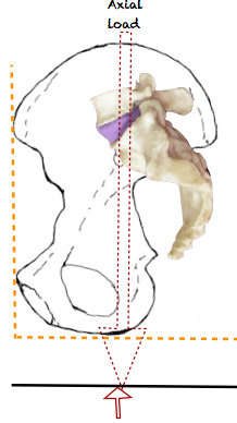
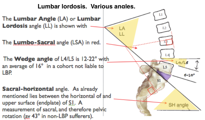
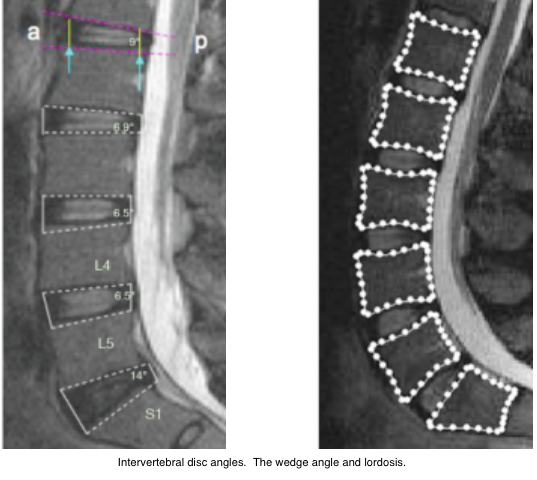
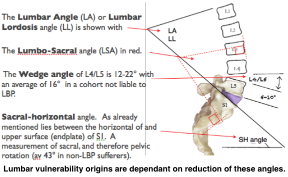
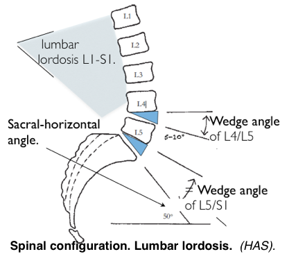
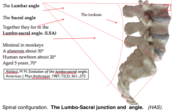
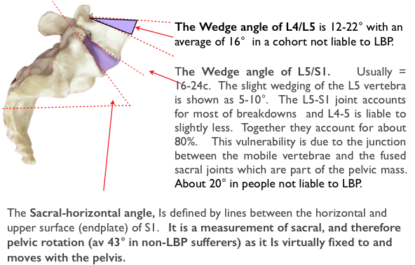
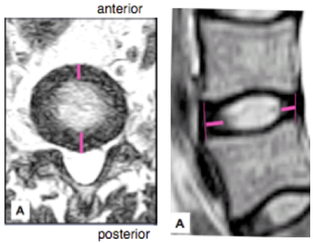
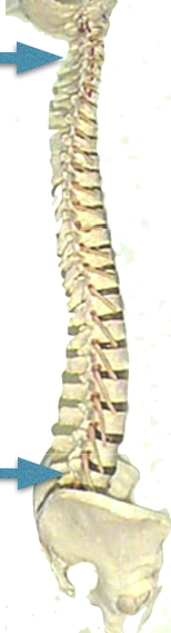
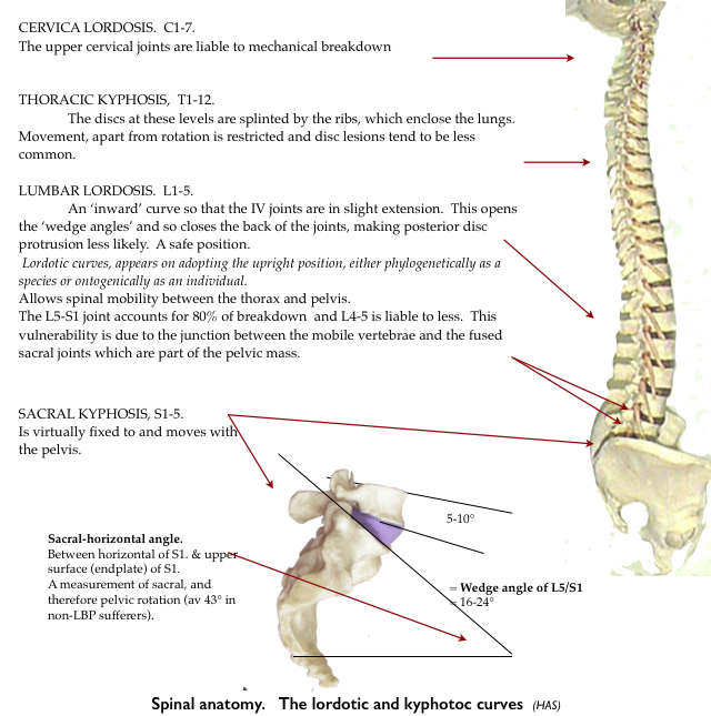
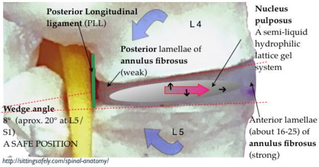
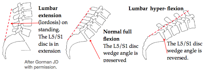
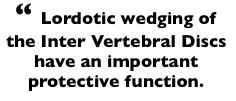
The orthograde upright spinal configuration was achieved, through natural selection, by the lordotic and kyphotic curves (below).   Lumbar vulnerability occurs when the wedge angle of the IV disc is reduced..

Lumbar vulnerability origins are dependant on reduction of these angles.

Lumbar vulnerability origins are dependant on reduction of these angles.

   The angles that determine lordosis have subsequently been extensively studied.   Note, in the diagram above, that the upper surface of S1 forms part of both the Sacral horizontal angle and the wedge angle of L5/S1.  The tilt of the pelvis therefore modifies L5/S1 angle.  Upright sitting effects the configuration and reduces the wedge angles.

Screen Shot 2015-05-31 at 16.12.10Already mentioned, the origins of lumbar vulnerability show that lordosis developed at two levels of the human spine, cervical and lumbar. Both these spinal levels are where  mobile segments meet a solid mass, the skull and the pelvis, and where mechanical spinal pathology mostly occurs and differences are found when comparing LBP patients with healthy patients (Jackson, 1994).   Cyriax wrote in 1946 that “the spinal joints subject to internal derangement are the 4th, 6th  & 7th cervical and the 4th & 5th lumbar”.       Cyriax also recognised that the lordotic wedging of the Inter Vertebral Discs (IVD) have an important function in protecting the discs (Harrison DD 1998) and is compromised by some sitting positions.Final pathway2

Natural Selection developed an upright (orthograde) posture  which results in :-

  • Lordotic changes to the lumbar spine to avoid walking with bent hips & knees (BHBK).   See below⟶
  • An increase of the IV Disk wedge angle.   This confers a degree of protection from NP retropulsion.
  • Rotation of the pelvic iliac blades for muscles to change from being extensors to abductors ensure pelvic stability.
  • Shortening of the ilium.
  • Relative reduction of the size of the birth canal.

Brain size

The early hominids, such as Homo erectus, had a brain of 900 cc. and its primitive variant, of 1.8 MYA, found at  the Dmanisi (Georgia) site was only 650-780cc. These were probably the earliest hominids outside of Africa (Lordkipanidze, 2005).    H. sapiens, with a volume of about 1300 cc appeared about 130,000 years ago according to the previous ‘out of Africa’ theory’ (Stringer 1970).

Bipedal rats and others 

 Hominids are the only known creatures which are truly bipedal and able to adopt our swinging gait apart possibly wingless birds, such as the ostrich, which are evolved from bipedal dinosaurs. The upright posture occurs in other animals but is usually for short periods and an examination of the skeleton, for example in the penguin, shows a different arrangement with only an analogous appearance of bipedalism.  Performing Japanese monkeys (Macaca fuscata).  can be trained to adopt an upright posture resulting in lumbar lordosis and bipedalism.  Over time some bone remodelling occurs.   However energy expenditure is higher than when plantigrade and they revert to this posture when retired from performing (Nakatsukas 2004).  Slijper, in 1942, gave a detailed account of the changes in the skeleton of a phocomelic goat that had  been born without forelegs. The spine and pelvis had been remodeled with changes suggestive of those found in bipedal animals.   

bipedal ratRats have been shown (Cassidy 1968) to adopt a bipedal stance and gait if their forelegs are amputated at birth. Their ability to function is remarkable. Their posture and locomotion are surprisingly similar to that of humans and provides the nearest animal model to the human bio-mechanical condition at the lumbar spine. The lumbar spine adapts by becoming lordotic and approximates to that of the human spine and there are changes in the muscles acting around the pelvis. It can be shown that there is increased axial loading on the lumbar spine and a high proportion of these rats develop back disorders which are usually, almost uniquely, only found in humans. These include degenerative changes, disc protrusion, facet joint degeneration and spinal stenosis.

Next for the obstetric pelvis  and it’s evolutionary importance ☛ see next post →